The heart and blood circulation, simply explained
Four chambers, four valves and two circuits: how the heart sends blood around the whole body in under a minute, and how to follow it step by step.
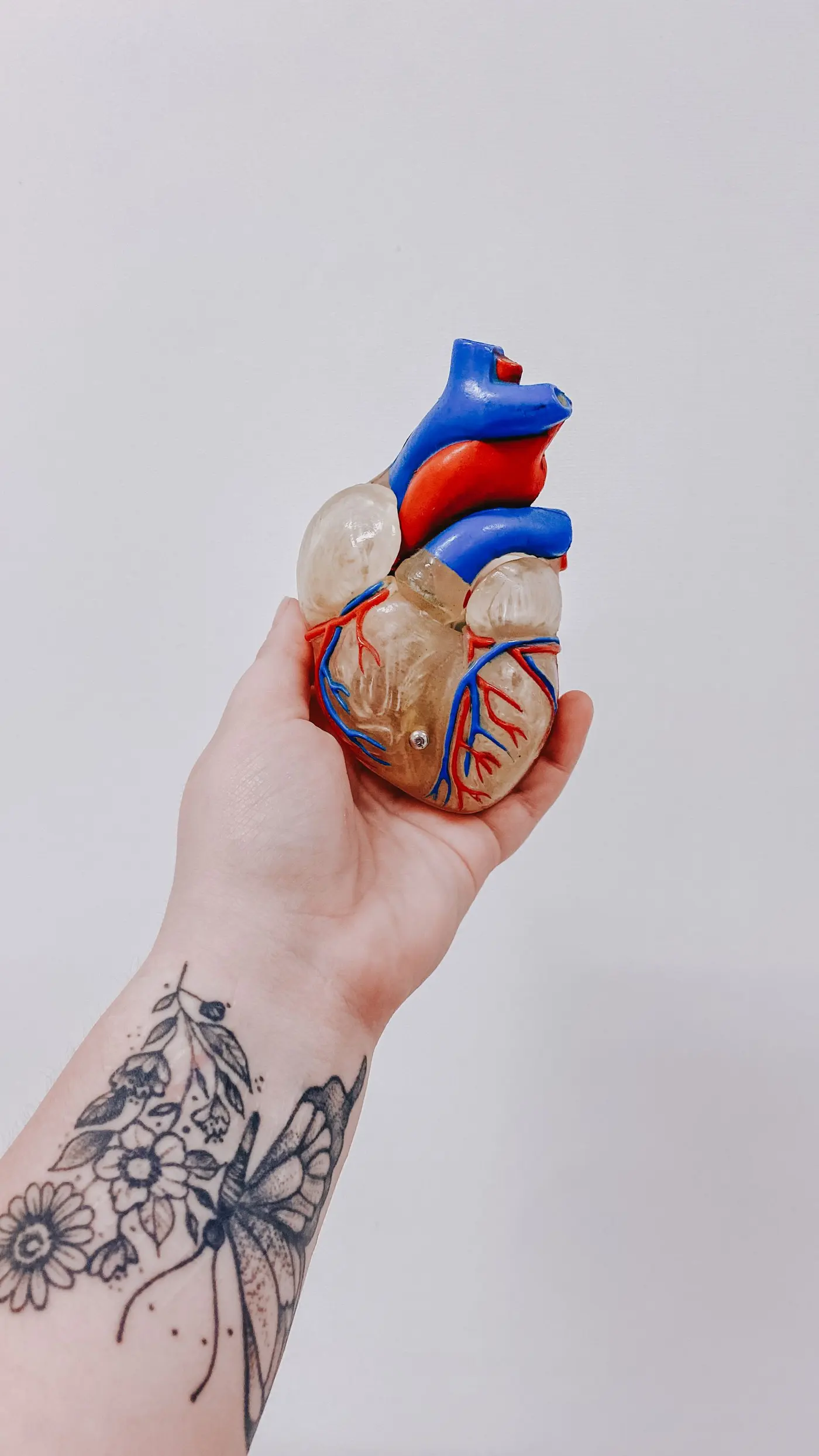
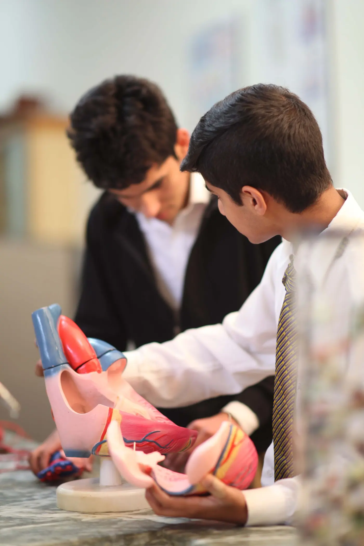
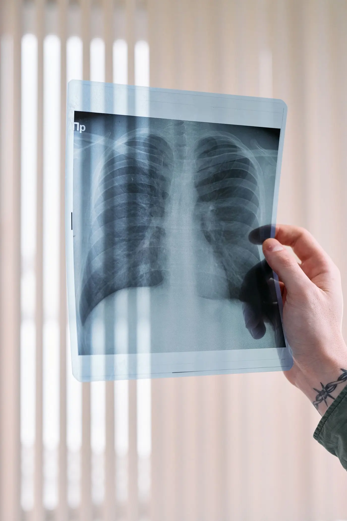
The heart is a hollow muscle about the size of a fist, sitting in the chest between the two lungs, slightly to the left. At rest, it beats around 60 to 80 times a minute, which adds up to more than 100,000 beats a day, without ever stopping.
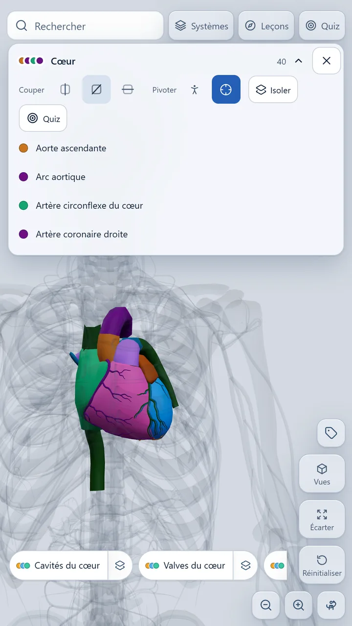
Four chambers, two pumps
The heart is divided into two halves that do not communicate. Each half has an atrium at the top, which receives blood, and a ventricle at the bottom, which pumps it out. The right heart sends blood to the lungs, the left heart to the rest of the body.
The left ventricle has the thickest wall: it is the one that has to push blood all the way to the tips of your toes.
Four valves for one-way traffic
Valves are flaps that stop blood from flowing backward. Between the atria and the ventricles sit the tricuspid valve on the right and the mitral valve on the left. At the exit of the ventricles are the pulmonary valve and the aortic valve. The "lub-dub" you hear through a stethoscope is the sound of them closing.
The path of blood, step by step
- 1Oxygen-poor blood returns from the body through the superior and inferior venae cavae.
- 2It enters the right atrium, then passes into the right ventricle.
- 3The right ventricle pumps it into the pulmonary artery, toward the lungs.
- 4In the lungs, it picks up oxygen and gets rid of carbon dioxide.
- 5It returns to the heart through the pulmonary veins, into the left atrium.
- 6The left ventricle propels it into the aorta, which distributes it to the whole body.
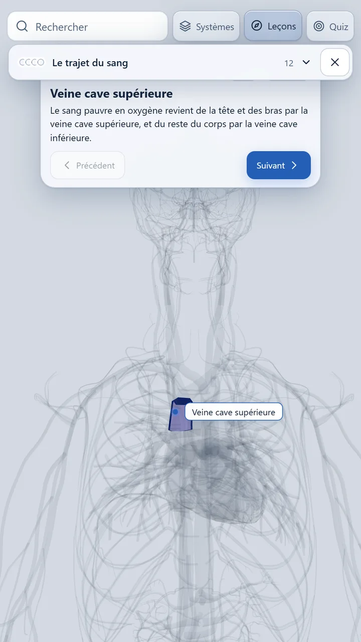
The heart needs feeding too
The heart muscle does not feed on the blood flowing through it: it gets its own supply from the coronary arteries, which branch off at the base of the aorta and run across its surface. When one of them gets blocked, part of the muscle is starved of oxygen: that is a heart attack (myocardial infarction).